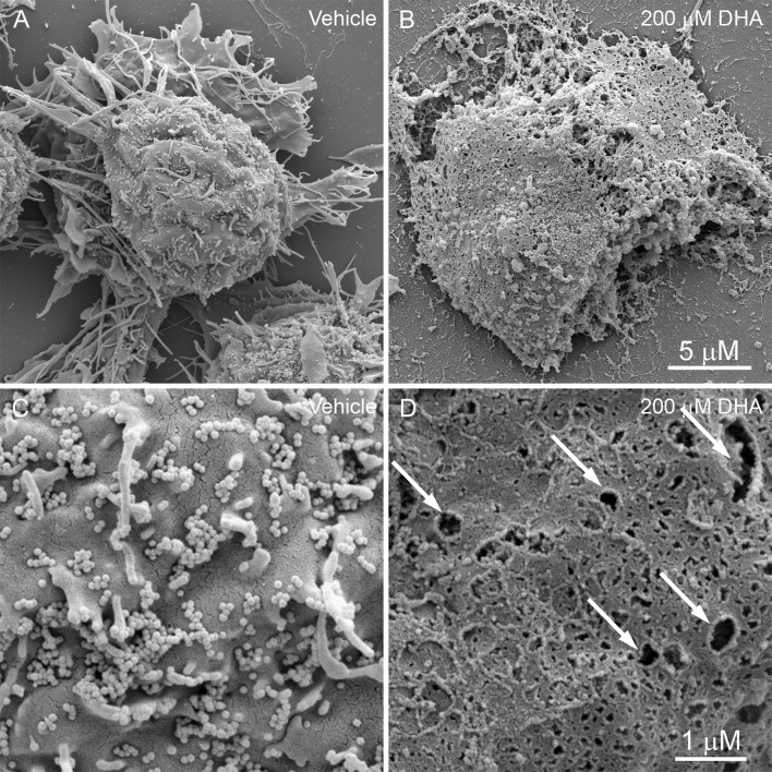
Fig. 2.
Scanning electron micrographs of BV-2 cell 4 h after treatment with vehicle (a, c) or 200 μM DHA (b, d). a At × 12,000 magnification, control cells are characterized by a rounded cellular mass with extended cellular processes. b DHA-treated cells have irregular margins and lack processes. The plasma membranes appear ruptured with extrusion of cellular contents. c At × 50,000 magnification, the surface of control cells exhibit projections from the plasma membrane and tethered extracellular vesicles (EVs). d DHA treatment causes the formation of membrane pits and pores of varying sizes, and a pronounced loss of EVs. Scale = 5 μm (a, b) and 1 μm (c, d)

