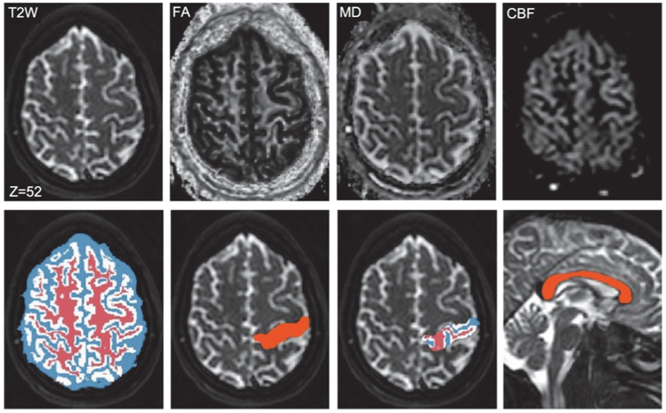
Fig. 3.
Sample parametric maps and label volumes in the space of T2W image of scan 1. Row one: Sample T2W image along with FA, MD, and CBF maps. Row two: Label maps of WM (red), GM (white), and CSF (blue); cross-section of a selected region in the parietal lobe; segmentation of the selected region into WM, GM, and CSF; and label map of the CC.

