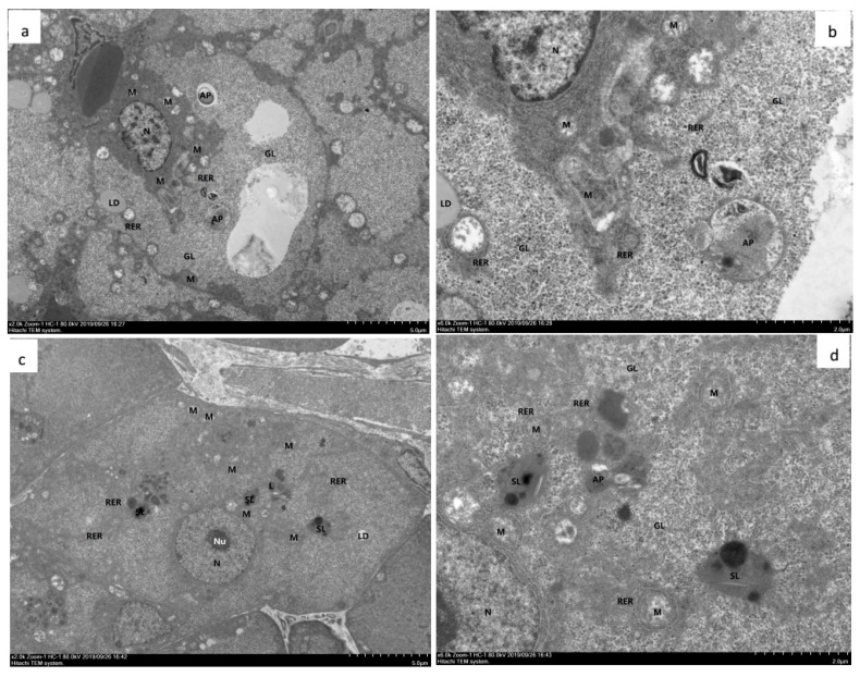
Figure 4.
Hepatopancreas from experimental diets treated with Litopenaeus vannamei. a–d TEM (a) and (b). The hepatopancreas from shrimp fed with OFO diet for 50 d. The hepatocytes showed moderate edema, intact cell membrane, moderate swelling and vacuolation of organelles in the cytoplasm, more vacuoles in the cells, and local electron density decreased. The mitochondria (M) showed slight swelling, most of the mitochondrial matrix slightly weakened, the internal cristae decreased, and the cristae disappeared in severe cases; the RER structure was fuzzy. Glycogen (GL) was abundant. Autophagy (AP) was abundant. Lipid droplets (LD) are present individually. There are more lysosomes (L) and secondary lysosomes (SL). (c) and (d) The hepatopancreas from shrimp fed with OFO/AX450 diet for 56 d. The liver cells showed mild edema, intact cell membrane, slight swelling of intracellular organelles, and a few vacuolations. autophagy (AP), mitochondria (M), large vacuoles (V), nucleoli (Nu), nucleus (N), Glycogen (GL), lysosomes (L), secondary lysosomes (SL), rough endoplasmic reticulum (RER).

