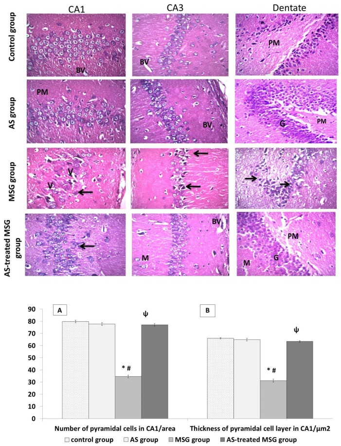
Figure 5.
Light microscopic examination of hematoxylin and eosin (H&E)-stained sections in all groups. Control and Allium sativum (AS) groups showed normal histological structure of hippocampus. Monosodium glutamate (MSG) group showed decrease in the number of the cells in Cornu Ammonis° (CA1 and CA3) areas with shrinkage and disarrangement of pyramidal cells. Apoptotic and pyknotic nuclei of pyramidal cells surrounded with pericellular haloes. Dilated blood vessels and massive vacuolation of the cytoplasm (V) can be also seen. Dentate gyrus of the same group showed many apoptotic cells (arrows), darkly stained nuclei, vacuolated cytoplasm (V), and disorganized blood vessels (BV). Allium sativum-treated MSG (AS-treated MSG) group was more or less like the control group, with multiple dilated blood vessels. (Hx. &E. X400). Histograms (A) and (B) indicate significant decrease in pyramidal cell count and layer thickness in the CA1 region in the MSG group compared to the control and AS groups. In addition, the AS-treated MSG group showed a significant increase in these parameters compared to the MSG group. * p < 0.05, vs. control; ψ p < 0.05, vs. MSG.

