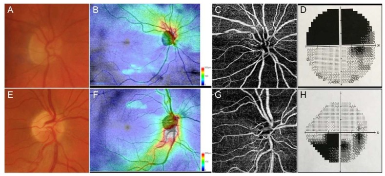
Figure 1.
Representative cases of anterior ischemic optic neuropathies. Anterior ischemic optic neuropathies (AAION) case showed a marked involvement of optic nerve head (A), with a strong damage of retinal nerve fiber layer (RNFL) (B), together with sectorial optical coherence tomography angiography (OCTA) perfusion impairment (C) and superior visual field defects (D). Non-anterior ischemic optic neuropathies (NAION) cases showed a less extensive optic nerve head (E) and RNFL (F) involvement, with a well-detected OCTA perfusion damage (G) and circumscribed visual field defect (H).

