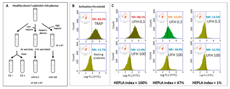
Figure 3.
Detection of heparin-dependent platelet-activating antibodies with FCA. (A) Schematic procedure of the FCA. (B) The first gate delineates the platelets ((log SSC + log FL2-PE (CD41+ events)) and the second gate is applied to analyze the activated platelets (log FL1-FITC). Determination of the activation threshold: upper panel, TRAP-activated platelets (Ctl+); lower panel, resting platelets (Ctl−), the cursor indicating the activation threshold is placed at the intersection of the FL1 histograms of the positive control (Ctl+) and the negative control (Ctl−). “%R” represents the percentage of CD-62P positive events as an index of platelet activation. This set-up allows determining percentage of the activated platelets (to the right of the activation threshold) under different conditions. (C) Incubations with patients’ plasma and one platelet donor: typical results with low (0.3 IU/mL heparin) and high (100 IU/mL heparin) concentrations of heparin (top and bottom rows, respectively). Left panels: platelets activated with a highly HIT-positive plasma (optical density, OD = 2.5); middle panels: platelets activated with a weakly HIT-positive plasma (OD = 1.3); right panels: platelets incubated with a non-HIT plasma. The HEPLA index is calculated as follows: HEPLA index = (% H 0.3 − % H 100)/(% TRAP Ctl+ − % PBS Ctl−) × 100. FCA: flow cytometric assay, SSC: sideways scatter, PE: phycoerythrin, FITC: fluorescein isothiocyanate, TRAP: thrombin receptor agonist peptide, UFH: unfractionated heparin.

