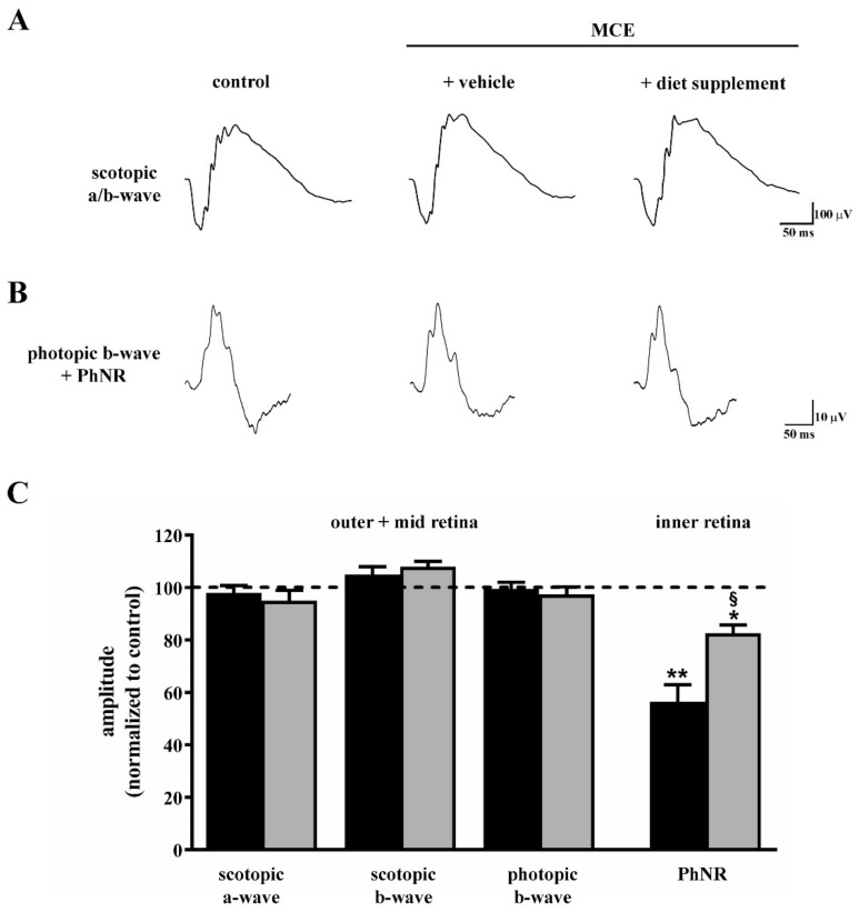
Figure 2.
Effects of dietary supplementation on retinal function as evaluated by scotopic and photopic full-field electroretinogram (ERG). Representative ERG traces showing scotopic a- and b-waves (A) or photopic b-waves with photopic negative response (PhNR; (B)) in control mice and in mice that received intraocular MCE injection fed with either vehicle or diet supplement. (C) Mean amplitudes of ERG responses evaluated as changes from baseline, normalized to the amplitude measured in control mice. MCE did not affect the amplitude of the scotopic a-wave, scotopic b-wave and photopic b-wave, while it reduced the amplitude of PhNR. Dietary supplementation partially prevented the reduction in PhNR amplitude. Data are shown as mean ± SEM (n = 6 for each group). * p < 0.01 and ** p < 0.001 versus control; § p < 0.001 versus MCE mice fed with vehicle (one-way ANOVA followed by the Newman–Keuls multiple comparison post-hoc test). Black bars: mice intraocularly injected with MCE fed with vehicle; grey bars: mice intraocularly injected with MCE fed with diet supplement.

