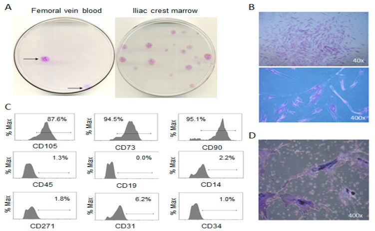
Figure 1.
Identification and phenotype of putative MSC colonies. (A) Macroscopic image of fibroblast-like colonies isolated from femoral vein blood (FVB) compared to iliac crest bone marrow, (B) microscopic images of FVB colonies, (C) histograms from flow cytometry of plastic adherent culture-expanded cells isolated from FVB (D) alkaline phosphatase staining of putative MSCs following osteogenic induction.

