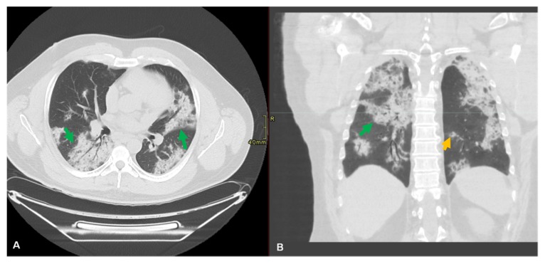
Figure 4.
Lung of a 51-year-old male patient with a history of hepatitis C and symptoms of dry cough and shortness of breathing for three weeks. No recent travel or known contacts with infected subjects. Axial (A) and coronal computed tomography (CT) (B) of chest without contrast revealed bilateral peribronchial and subpleural consolidative opacities noted throughout both lungs (green arrow). There were scattered nodular consolidative opacities in a peribronchial distribution (orange arrow). The patient tested positive for SARS-CoV-2 RNA.

