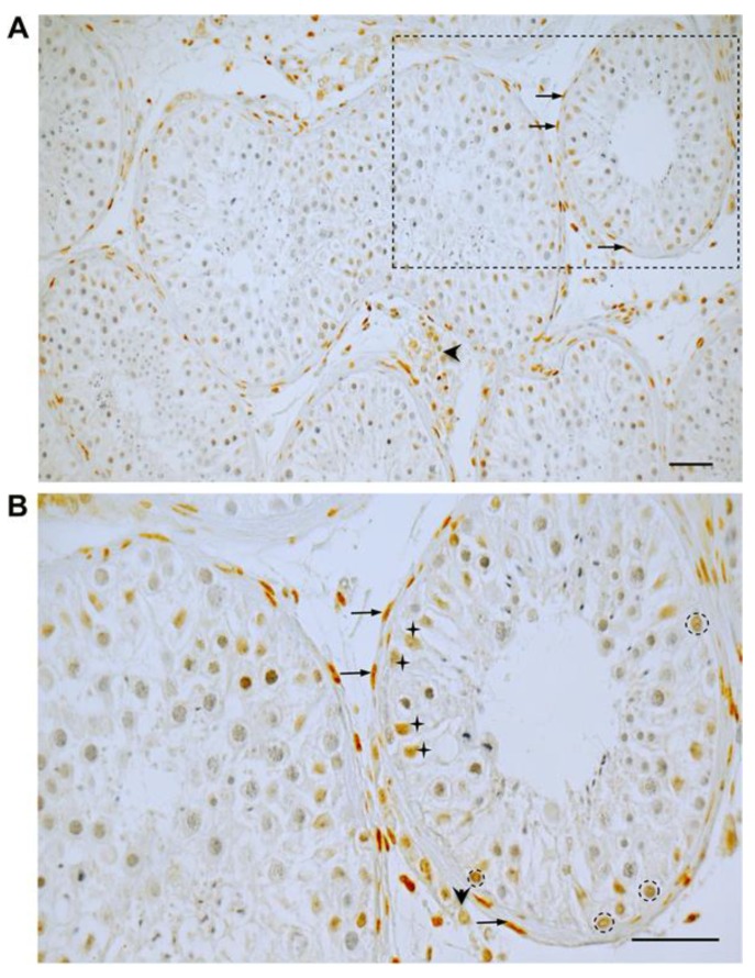
Figure 1.
Immunohistochemistry of GR in the human testis (A,B = boxed area in A). In a representative human testicular biopsy, GR staining is found in peritubular, myoid cells (arrows in A,B) building the wall of seminiferous tubules, in some Sertoli cells (crosses, B) and some spermatogonia (circles, B). In addition, various interstitial cells, presumably Leydig cells (arrowheads in A,B), but also macrophages and endothelial cells of blood vessels, are immuno-positive. Nuclei were slightly stained with Hematoxylin. Scale bars = 50 µm.

