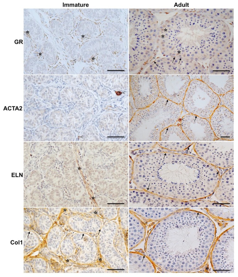
Figure 2.
Expression of GR, ACTA2 and extracellular proteins in peritubular cells of the monkey testis before and after puberty. Immunohistochemistry of GR of adult rhesus monkeys (right panels) shows strong GR expression in the peritubular compartment (arrows) and moderate staining of Sertoli cells (asterisks). Smooth muscle cells of blood vessels (arrowheads) served as a positive intrinsic control. GR staining of the peritubular wall was not found in all samples from immature rhesus monkeys (186–282 days, left panels and results not shown). Only interstitial cells (asterisks) and blood vessels (arrowheads) show a robust staining. ACTA2 is constantly expressed by vascular smooth muscle cells (arrowheads) in both immature and adult monkeys but only localized in the peritubular wall of mature monkeys (arrows). In immature rhesus, staining of ELN is restricted to the interstitial matrix (asterisks) but is not detected in the peritubular compartment, which in turn is the prevailing expression site in adult monkeys (arrows). Independent of age, Col1 is localized in the peritubular compartment (arrows), in the interstitial area (asterisks), and around blood vessels (arrowheads). Negative control experiments were performed with IgG (not shown). Hematoxylin was used to counterstain nuclei. Scale bars = 50 µm.

