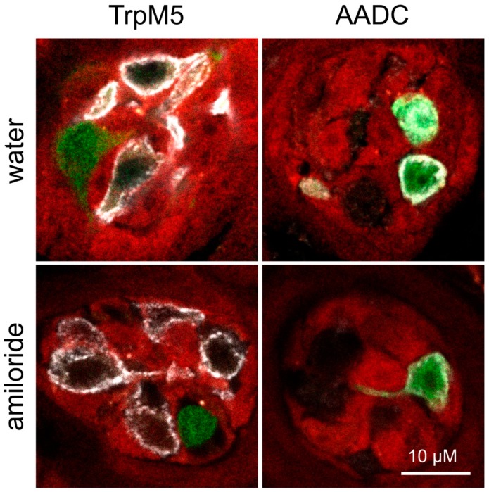
Figure 4.
Expression of fluorescent proteins in taste papillae after amiloride intervention. Fungiform papillae sections of Scnn1aa/bb animals expressing GFP (green) and tdRFP (red) fluorescence in αENaC- and βENaC-expressing cells, respectively, were stained for Type II (TrpM5) and Type III (AADC) taste cell markers after amiloride intervention. Therefore, animals received adequate salt diet without or with 300 µM amiloride-containing drinking water prior to sacrifice. Independent of intervention, GFP and tdRFP fluorescence showed no co-localization in taste papillae. Whereas GFP-positive cells always co-expressed AADC, tdRFP-positive cells revealed no overlap with the cell markers TrpM5 or AADC, visualized by immunofluorescence (white). Scale bar applies to all images.

