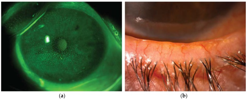
Figure 1.
Representative image from a patient with dry eye disease owing to Meibomian gland dysfunction. (a) Slit lamp photograph of the cornea after instillation of 20 µL unpreserved 2% sodium fluorescein (corneal fluorescein staining). The epithelial damage is visible as multiple punctate epithelial erosions staining with fluorescein scattered over the corneal surface. (b) Slit lamp photograph of the lid margin showing typical signs of meibomian gland dysfunction, including hyperemia and irregularity of the lid margin, telangectasia, plugged gland orifices and anterior displacement of the mucocutaneous junction. Hyposecretion of meibomian lipids leads to tear film instability and increased evaporation rate.

