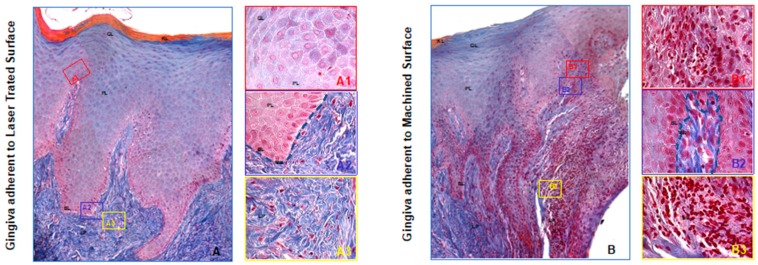
Figure 3.
Trichrome staining of the region of the gingiva adherent to the laser-conditioned- (A) or to machined surfaces (B). In higher magnification of the gingiva adherent to laser treated surface it is evident the regular morphology in all layers: Junctional epithelium (A1), sulcular epithelium with all the typical layers including continuous basal membrane highlighted with the dotted blue line (A2) and subepithelial connective tissue of the lamina propria (A3). In higher magnification of the gingiva adherent to machined surface the typical morphology of the junctional epithelium is not respected (B1): the basal membrane is disrupted (highlighted with the dotted blue line in (B2) and the collagen fibers of subepithelial connective tissue of the lamina propria looks infiltrated with large inflamed area (B3). Magnification 10X (A,B) or 40X (A1–3,B1–3). Red, yellow and blue rectangles in A and B represent enlarged areas in the panels on the side, as indicated. Panel A and B are the results of sequential images mounted together. LP: Lamina propria; MB: basilar membrane (dotted blue line); BL: Basal Layer; PL: Prickle layer; GL: granular layer, KL: keratinized layer.

