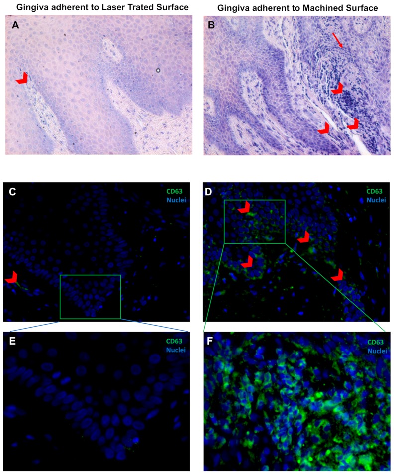
Figure 5.
May–Grünwald–Giemsa staining (A,B) and Immunofluorescence against CD63 (green) (C,D) of the soft tissue area adherent to the laser-treated (A,C,E) or to machined (B,D,E) surfaces, as indicated. Red arrowheads indicate flogistic area with leukocites accumulation (A,B), that are also positive to CD63 immunostaining (C–F). Nuclei are counterstained with 4’,6-diamino-2-phenylindole DAPI in (C,F). Magnification 10X (A,B), 20X (C,D) or 40X (E,F).

