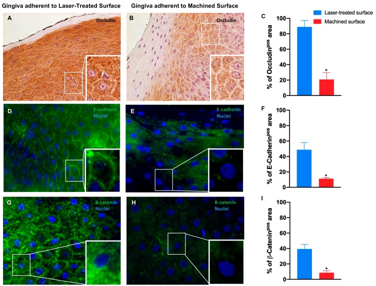
Figure 7.
Immunostaining against occludin (A,B) and immunofluorescence for E-cadherin (D,E) and β-catenin (G,H) of the region of gingiva adherent to the laser-treated surface and to machined surface, as indicated. Magnification: 20X (or 40X in the inserts) in (A,B); 40X and (or 100X in the inserts) in (D,E,G). Percentage of occludin (C), E-cadherin (F) and β-catenin (I) positive staining area in the epithelial layers are pointed out in the bar graphs. Values are expressed as mean ±SD of five determinations in randomly chosen sections for each specimen. The (*) indicates values statistically different (p < 0.001) between the area of the soft tissue’s adherent to the laser-treated- or to machined surfaces, as indicated.

