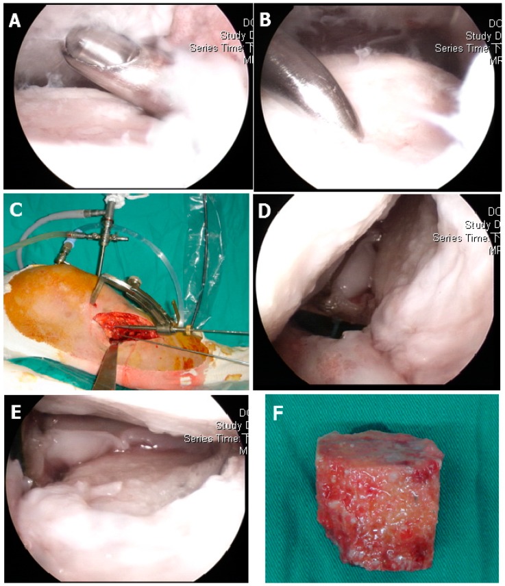
Figure 1.
Operative procedure: (A) Arthroscopic documentation and debridement. (B) Use of a microfracture awl to identify the intra-articular depression zone. (C) Anterior cruciate ligament (ACL) tibial guiding for osteotomy axis and location, where two parallel Kirschner wire determined one plane, the 5 mm osteotome started from the metaphysic area to the articular surface. (D) The malunited fracture area was completely released. (E) The depressed articular fragments were elevated with bone tamp under direct arthroscopically inspection. (F) Bone grafting of the metaphyseal defect with structural allograft femoral head.

