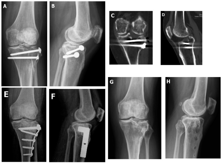
Figure 2.
(A,B) This 43-year-old patient had a Type II fracture, where open reduction with internal fixation was done by two cannulated screws on the day of injury. This radiograph was checked four months after the initial surgery, where a lateral split with depression was noted. (C,D) CT revealed lateral plateau huge intraarticular split and depression. (E,F) Immediate postoperative radiograph after arthroscopically-assisted corrective osteotomy (AACO), bone grafting with structural allograft femoral head, and internal fixation by lateral plate. (G,H) 4 years after AACO, the plate was removed and the radiological score were excellent.

