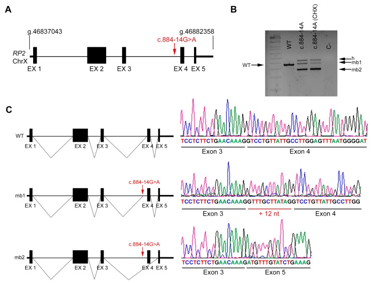
Figure 4.
In vivo splicing analysis of the non-canonical splice site (NCSS) variant identified in RP2. (A) Diagram showing the genomic position of the NCSS variant in intron 3 of the RP2 gene in chromosome X (c.884 -14G>A) from patient R1. (B) In vivo analysis of RP2 mRNAs from control (WT) and patient’s blood samples (treated or untreated with cycloheximide, CHX). The band of 370 bp corresponds to the wild-type transcript (WT), whereas blood from the hemizygous patient carrying the NCSS variant resulted into two different aberrant transcripts (mb1 and mb2). h indicates the heteroduplex band of mb1 and mb2. C- indicates the PCR negative control. (C) Subsequent Sanger sequencing of cloned individual bands, indicated in the left diagrams, confirmed the in-frame insertion of 12 bp (mb1) and exon skipping of exon 4 (mb2) compared to the wild-type transcript.

