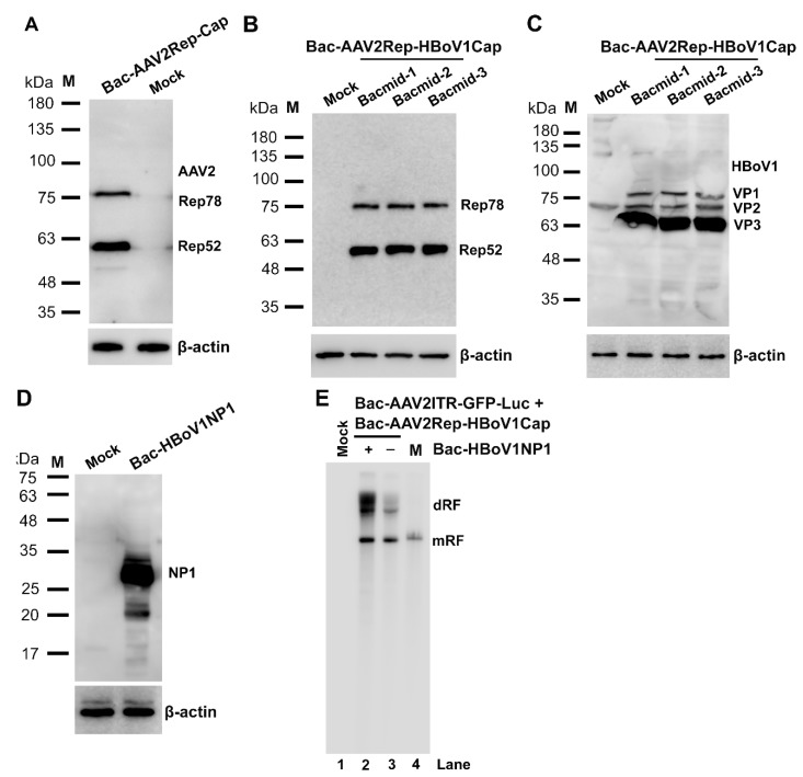
Figure 2.
Expression of AAV2 Rep and Cap proteins and HBoV1 Cap and NP1 proteins in BEV-infected Sf9 cells. (A–D) Western blotting. Sf9 insect cells were infected with Bac-AAV2Rep-Cap (A), Bac-AAV2Rep-HBoV1Cap (B,C), or Bac-HBoV1NP1 (D). The infected cells were collected at 72 hrs post-infection and subjected to Western blot analysis. (A) AAV2 Rep proteins were detected with an anti-Rep monoclonal antibody. (B,C) Bac-AAV2Rep-HBoV1Cap generated from transfection of three bacmid (Bacmid-1-3) were infected with Sf9 cells independently. (B) AAV2 Rep proteins were detected with an anti-Rep monoclonal antibody, and (C) HBoV1 Cap protein expression was detected with an anti-HBoV1 Cap protein antiserum. (D) HBoV1 NP1 was detected with a rat anti-HBoV1 NP1 antiserum. β-actin served as a loading control. Mock, uninfected cells. (E) Southern blotting. Sf9 cells were infected with Bac-AAV2ITR-GFP-Luc and Bac-AAV2Rep-HBoV1Cap with (+) or without (-) co-infection of Bac-HBoV1NP1. Cells were collected at 72 hrs post-infection and subjected to extraction of lower molecular weight (Hirt) DNAs, which were analyzed by Southern blotting. Mock, uninfected Sf9 cells as a control; M, a marker of a rAAV2ITR-GFP-Luc proviral replicative form (RF) genome of 5.4 kb. dRF and mRF, double and monomer RF.

