Figure 2.

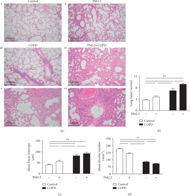
The effect of PM2.5 on lung histological injury in rats with COPD. (a) Lung tissue HE staining of different groups of rats. (i) Control, an intact structure of pulmonary alveoli and airway (magnification, ×100); (ii) PM2.5, mild infiltration of inflammatory cells and thickening of airway wall (magnification, ×100); (iii) COPD, alveolar cavity enlargement and vascular wall thickening (magnification, ×100); (iv) PM2.5+COPD, chronic obstructive bronchiolitis with thickening of the airway wall and infiltration with inflammatory cells (magnification, ×100); (v) COPD, airway wall thickening and inflammatory cell infiltration (magnification, ×200); (vi) PM2.5+COPD, increased thickening of airway wall and infiltration of inflammatory cells compared to COPD (magnification, ×200). (b–d) Quantitative analysis of lung injury, that is, the level of (b) LIS, (c) MLI, and (d) MAN of the lungs in each group. LIS: lung injury scores; MLI: mean linear intercept; MAN: mean alveolar number. The data are expressed as the means ± SD (n = 6~7). ∗∗p < 0.01, ∗p < 0.05.
