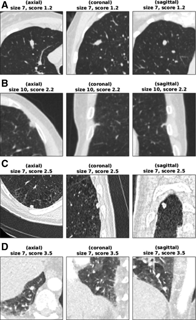
Figure 3.
Low-scoring cancer cases. (A) Woman aged 61 years (smoking status: ex-smoker) with a 7 mm cancer located in RUL, scoring 1.19 (Brock=3.50). The median HU value in the aortic arch is 37. (B) Man aged 61 years (smoking status: unknown) with a 10 mm cancer located in the lingula lobe, scoring 2.18 (Brock=5.83). The median HU value in the aortic arch is 135. (C) Man aged 67 years (smoking status: current smoker) with a 7 mm cancer located in RLL, scoring 2.55 (Brock=1.31). The median HU value in the aortic arch is 50. (D) Woman aged 71 years (smoking status: unknown) with a 7 mm cancer located in RLL, scoring 3.46 (Brock=2.26). The median HU value in the aortic arch is 217. CT appears not to be using a breath-hold protocol. The only cancer actually stratified into the ‘rule-out’ set is (A), possibly because of its atypical shape and smooth appearance. The cancer in (B) was not reimaged for another 2 years after this scan, and the patient’s lungs had several similar lesions that did not grow into cancers. For cases such as (D), reimaging the nodule with a standard breath-hold protocol would be expected to give a cleaner image on which the lung cancer prediction convolutional neural network yields a higher score. HU, Hounsfield unit; RLL, right lower lobe; RUL, right upper lobe.

