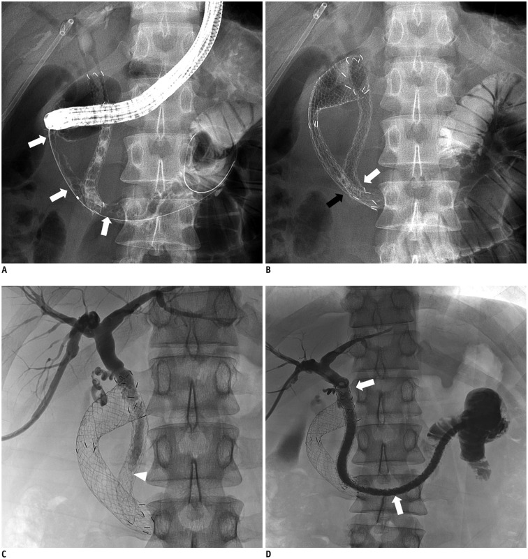
Fig. 3. 31-year-old man with pancreatic cancer. Biliary obstruction in this patient had been treated with covered biliary stent 2 weeks previously.
A. Fluoroscopic image showing type II duodenal stricture (white arrows). Contrast media had refluxed into previously inserted biliary stent. B. Placement of an uncovered duodenal stent (20 mm × 10 cm). Overlap of distal ends of duodenal stent (black arrow) and biliary stent (white arrow) is evident. C. Cholangiogram via right PTBD at one week after duodenal stent placement indicating stasis of contrast medium in common bile duct (arrowhead), suggesting biliary stent malfunction due to extrinsic compression by subsequently inserted duodenal stent. D. Additional long type GD stent (10 mm × 23 cm, white arrows) was deployed through lumen of previous biliary stent into proximal jejunum. Cholangiogram showing good stent position and expansion, as well as good passage of contrast medium to jejunum via subsequent stent.

