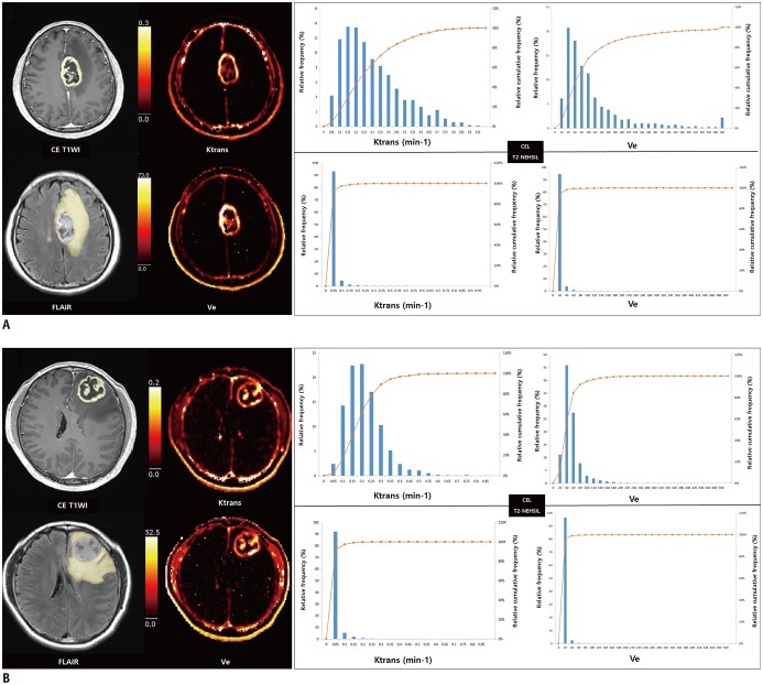
Fig. 3. Representative dynamic contrast-enhanced MR imaging-derived pharmacokinetic variable maps in patients whose GBM had progressed (A) and had not progressed (B) after standard treatment.
A. 53-year-old GBM patient who had early disease progression after standard treatment (PFS = 9 months). Preoperative transverse CE T1WI and FLAIR images show ROI of CEL and surrounding NE-T2HSIL, respectively. Preoperative pharmacokinetic DCE parametric maps of Ktrans and Ve in CEL show higher values as compared with those of surrounding NE-T2HSIL. B. 43-year-old GBM patient who did not progress after standard treatment (PFS = 55 months). On histograms for pharmacokinetic variables, lines represent relative cumulative frequencies of Ktrans and Ve. Histograms of CEL of patient (B) show rightward shift as compared with corresponding histograms in (A), suggesting that frequencies of low values were higher in patients who had not progressed than patients with early disease progression. PFS = progression-free survival

