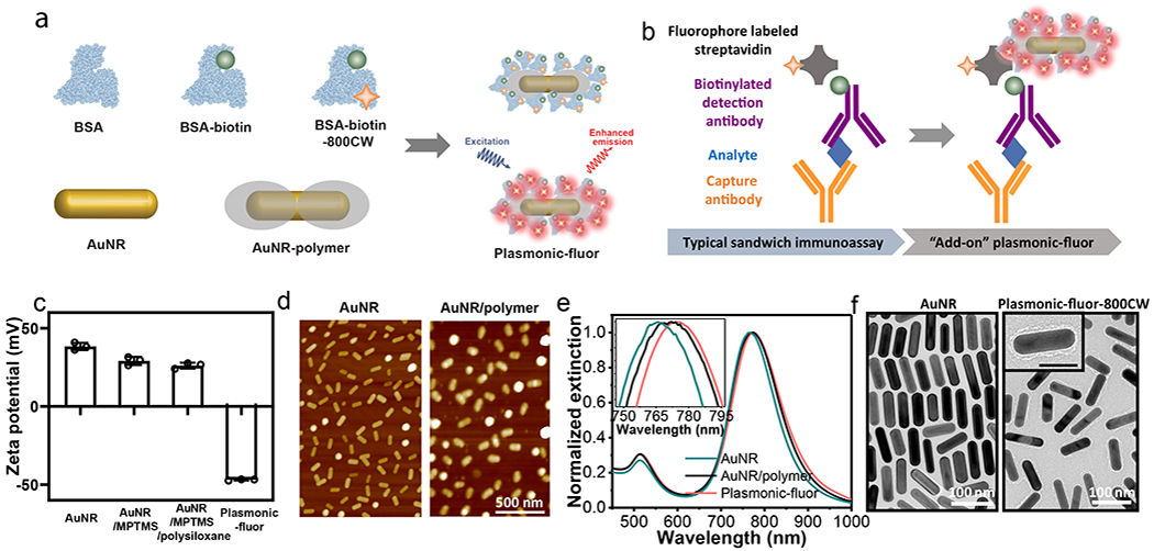
Fig. 1 |. Plasmonic-fluor synthesis and material characterization.

a, Schematic illustration showing the structure of “plasmonic-fluor”, which consists of a plasmonically-active core (e.g. gold nanorod (AuNR)), a polymer shell as spacer layer, light emitters, and a universal biorecognition element (biotin). Bovine serum albumin (BSA) is employed as a key design element to assemble all components into the functional nanoconstruct and to resist non-specific binding. b, Working principle of plasmonic-fluor as an “add-on” biolabel to enhance the fluorescence intensity and consequent signal-to-noise ratio of fluorescence-based assays, without changing the existing assay workflow. c, Zeta potential of AuNR, AuNR/MPTMS, AuNR/MPTMS/polysiloxane (AuNR/polymer), and the plasmonic-fluor-800CW (AuNR/polymer/BSA-biotin-800CW). Error bar, s.d. (n=3 repeated tests). d, AFM images showing the AuNR before and after coating with polymer. e, Vis-NIR extinction spectra of AuNR, AuNR/polymer, and plasmonic-fluor, showing a progressive red shift in the LSPR wavelength after each step. f, TEM images of bare AuNR and plasmonic-fluor-800CW.
