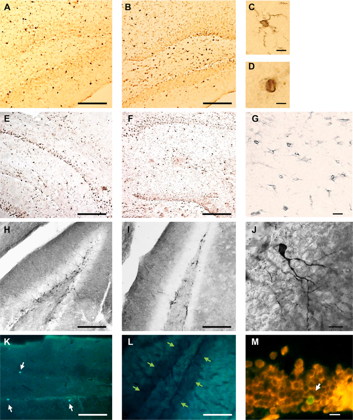
Fig. 3.
Representative IHC. Iba-1+ cells in A. VEH-treated and B. TX-treated mouse, C. Ramified, and D. Amoeboid microglia in a TX-treated mouse. E. IL-1β + cells in VEH-treated and F, G, TX-treated mice. H. DCX+ cells in the dentate gyrus of VEH-treated and I. NTX-treated mouse. J. DCX+ cell from H. K. DG viewed under FITC filter showing 3 BrdU+ cells in VEH-treated mouse (white arrows) and L. lack of BrdU+ cells in TX-treated mouse. Green arrows indicate the granular layer of the dentate gyrus. M. BrdU + /NeuN+ cell (white arrow) in the granule cell layer of the DG shown with a double FITC/rhodamine filter in VEH-treated mouse. All sections are dorsal hippocampus. Scale bars A, B, E, F, H, I, K, L 250 μm; C,D = 10 μm; G = 25 μm; J,M = 20 μm. A, B, E, F, H, I, K, L viewed under 10x magnification; C, D, G, J, M, viewed under 60x magnification.

