Figure 1.
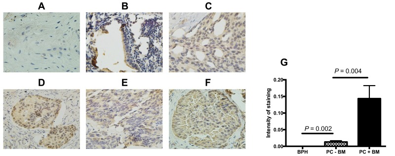
BTK protein expression in the tissues of prostate cancer and benign hyperplasia. BTK protein expression was assessed by immunohistochemistry as described in the methods. (A) Benign prostatic hyperplasia; (B) prostate cancer without bone metastasis and Gleason score 7 (3+4); (C) prostate cancer with bone metastasis and Gleason score 7 (4+3); (D) prostate cancer with bone metastasis and Gleason score 8 (3+5); (E) prostate cancer without bone metastasis and Gleason score 8 (4+4); (F) prostate cancer with bone metastasis and Gleason score 9 (5+4). Magnification: ×400 for A, C, E, and F; ×200 for B and D. (G) Semi-quantitative comparison of the immunostaining intensity. Vertical axis: Intensity of staining (orbital value obtained by the imaging processing software), horizontal axis: groups of the samples.
Abbreviations: BPH, benign prostatic hyperplasia; PC-BM, prostate cancer without bone metastasis; PC+BM, prostate cancer with bone metastasis.

