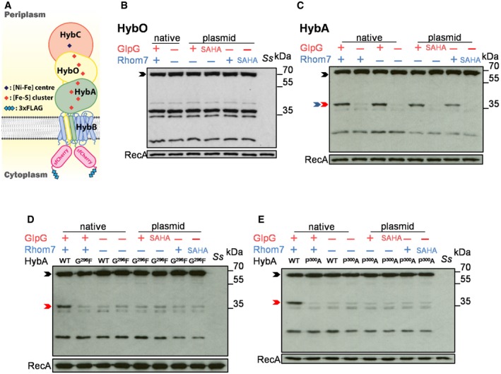
Schematic of HybA and HybO fusions.
Western blot analysis (probing with an anti‐FLAG mAb) to detect cleavage of HybO with (+)/without (−) chromosomal (native) or pBAD33‐encoded rhomboids (plasmid).
Western blot analysis to detect cleavage of HybA with (+)/without (−) chromosomal (native) or pBAD33‐encoded rhomboids (plasmid).
Western blot analysis to detect cleavage of HybAG296F by endogenous or pBAD33‐encoded rhomboid.
Western blot analysis to detect cleavage of HybAP300A by endogenous or pBAD33‐encoded rhomboid.
Data information: In (B‐E), rhomboid substrates that are uncleaved, cleaved by GlpG or cleaved by Rhom7 are marked by black, red and blue arrows, respectively. Wild‐type
Shigella sonnei,
Ss. Wild‐type (WT)/inactive (SAHA: alanine substitution of the catalytic serine and histidine residues) enzymes were pBAD33‐encoded (plasmid) in
S. sonnei Δ
glpGΔ
rhom7. RecA, loading control.
Source data are available online for this figure.

