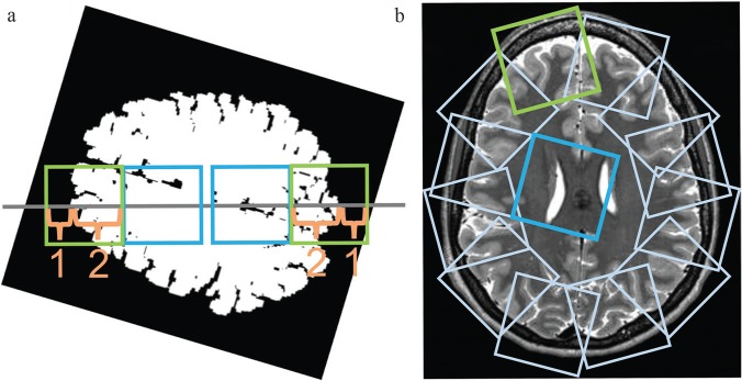
Fig. 1.
Twelve subimages from around the periphery of the brain and another 12 subimages from the inner area of the brain were subsampled automatically from each slice selected in preprocessing step 3. (a) First, each slice image was rotated through a random angle around the center of the brain. Four small subimages (64 × 64 pixels each) were then defined on a horizontal line passing through the center of the brain (gray line). The first two images were taken from the peripheral brain area (green squares), with the ratio of the brain parenchyma length to the extra-parenchyma length along the line being 2:1. Next, another two subimages (64 × 64 pixels each, blue squares) adjacent to the peripheral images on the medial sides were selected as images from the inner area of the brain. (b) The subsampling of peripheral and medial images was repeated 12 times after rotating the line in 30° increments beginning from the initial orientation. Overall, the procedure results in 12 subimages sampled at regular angular intervals for both the peripheral and the inner area of the brain.

