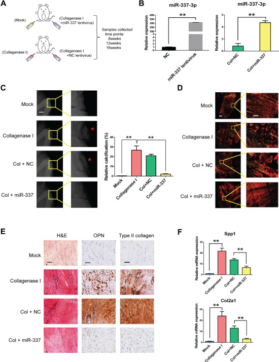
Figure 2.

miR-337-3p counteracts chondro-osteogenesis in collagenase I-induced rat tendinopathy model. (A) Schematic diagram of applying rno-miR-337-3p overexpressing lentivirus to cure rat tendinopathy induced by collagenase I. All reagents were injected at patellar tendon. Male SD rats (8-week-old) were used for the experiment, and patellar tendon samples were collected after 8, 12, and 16 weeks. Mock group stands for saline injection and suture. (B) Real-time PCR analysis of miR-337-3p in rTDSCs transfected with miR-337-3p overexpressing lentivirus for 7 days (left) and in patellar tendon tissues of rat tendinopathy model treated with miR-337-3p overexpressing lentivirus (right). (C) X-ray image of the knees of SD rats at 12 weeks after treatment showed that knee joints and ectopic ossicles (red asterisks in the magnified pictures) formed in patellar tendons (left). Scale bar, 5 mm. Relative calcification percents were measured through X-ray results by Image J (right). (D) Patellar tendon paraffin sections collected from each group at 12 weeks were treated with Sirius red staining and observed under polarized light microscopy to observe the collagen fibers. Left columns are images of integrated intact patellar tendon. Right columns are enlarged partial patellar tendon images. Scale bar, 800 μm (left) and 200 μm (right). (E) H&E staining and immunocytochemistry staining of OPN and type II collagen in each group. Samples were collected at 12 weeks after surgery. Scale bar, 50 μm. (F) Real-time PCR analysis of Spp1 and Col2a1 in patellar tendon tissues of each group. Error bars, SEM (n = 3). **P < 0.01.
