Figure 1.
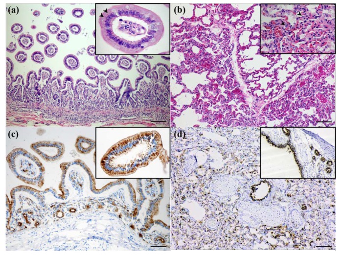
Civet tissue staining. (a,b); H&E staining showing (a) severe chronic lymphoplasmacytic enteritis with eosinophilic intranuclear and intracytoplasmic inclusion bodies that are frequently seen within villous epitheliums (inset, arrowheads) and (b) severe broncho-interstitial pneumonia with eosinophilic intracytoplasmic inclusion bodies that were randomly observed in pneumocyte-lining alveoli (inset, arrowheads). (c,d), IHC staining showing the immunopositive CDV antigen was diffusely observed within (c) cryptal and villous epitheliums (inset) and (d) in pulmonary and bronchial epitheliums (inset). Scale bar = 200 µm.

