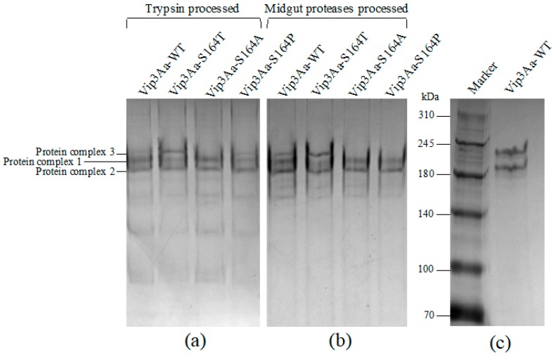
Figure 2.
Analysis of native Vip3Aa proteins after proteolytic processing. Protease treated Vip3Aa-WT, Vip3Aa-S164T, Vip3Aa-S164A and Vip3Aa-S164P by either commercial trypsin or midgut proteases of S. litura larvae were analyzed by the electrophoretic analysis without heat denaturation. (a) Vip3Aa proteins after tryptic processing were analyzed in a native gel; (b): Vip3Aa proteins after processing by midgut proteases were analyzed in a native gel; (c): Vip3Aa proteins after tryptic processing were mixed with 5 × SDS-PAGE sample buffer without β-mercaptoethanol and analyzed in an SDS-PAGE gel. Protein complex 1, protein complex 2 and protein complex 3 in panel (a) indicate gel bands sliced from each lane in the native gel.

