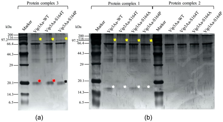
Figure 3.
Separation of peptides from protein complexes of tryptic Vip3Aa proteins. Major protein bands representing different protein complexes in Figure 2a were sliced and separated in an SDS-PAGE gel. (a) peptides separated from the protein complex 3 in Figure 2a; (b) peptides separated from the protein complexes 1 and 2 respectively in Figure 2a. The yellow, red, black and white arrows indicate the bands of 95 kDa, 19 kDa, 17 kDa and 15 kDa fragments respectively.

