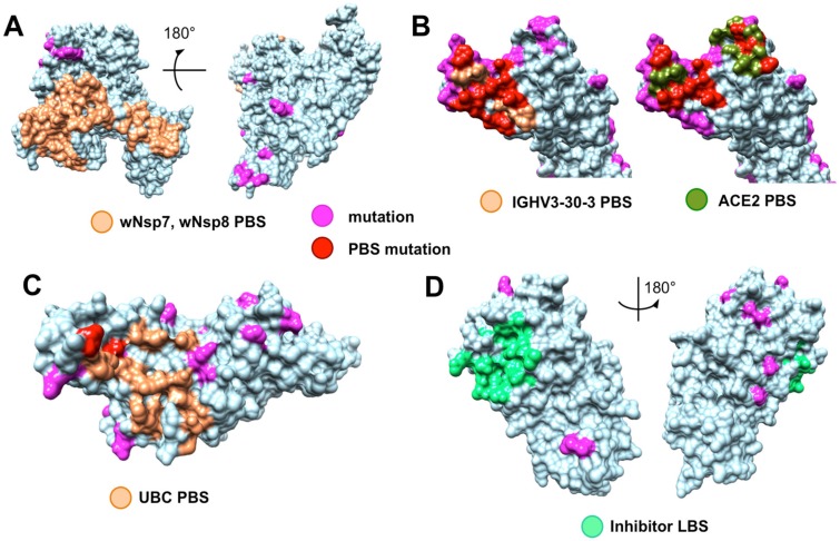
Figure 4.
Evolutionary conservation of functional sites in SARS-CoV-2 proteins. (A) Fully conserved protein binding sites (PBS, light orange) of wNsp12 in its interaction with wNsp7 and wNsp8, while other parts of the protein surface shows mutations (magenta); (B) Both the major monoclonal antibody binding site (light orange) and the ACE2 receptor binding site (dark green) of wS are heavily mutated (binding site mutations are shown in red) compared to the same binding sites in other coronaviruses; mutations not located on the two binding sites are shown in magenta; (C) Nearly intact protein binding site (light orange) of wNsp (papain-like protease PLpro domain) for its putative interaction with human ubiquitin-aldehyde (binding site mutations of the only two residues are shown in red, non-binding site mutations are shown in magenta); (D) Fully conserved inhibitor ligand binding site (LBS, green) for wNsp5; non-binding site mutations are shown in magenta.

