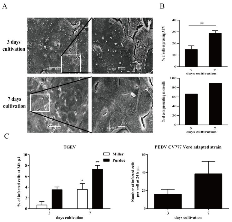
Figure 1.
Effect of cultivation age on the percentage of microvilli positive cells and susceptibility to virus infection. (A) At three days and seven days cultivation, the microvilli on the surface of primary enterocytes were detected by scanning electron micrograph. (B) The percentages of microvilli positive cells and aminopeptidase N (APN) positive cells were counted. (C) At three days and seven days cultivation, cells were inoculated with Porcine epidemic diarrhea virus (PEDV; 105.6 TCID50/mL) and transmissible gastroenteritis virus (TGEV; m.o.i. = 1). Percentage of infection was measured 24 h post inoculation. Data are expressed as mean ± standard deviation (SD) of the results of three separate experiments. Statistically significant differences in comparison with data from three days cultivation are presented as *p < 0.05 or **p < 0.01.

