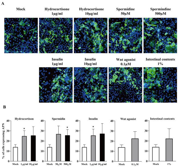
Figure 2.
APN expression in primary ileum epithelial cells in the presence of enterocytes differentiation factors hydrocortisone, spermidine, porcine insulin, Wnt agonist, or small intestinal contents. (A) Immunofluorescence staining of APN expression (green) in enterocytes with different treatments. Scale bar: 50 µm. (B) The percentage of cells expressing APN at 24 h of treatment. Data are expressed as mean ± SD of the results of three separate experiments. Statistically significant differences in comparison with data from mock treatment are presented as *p < 0.05.

