Figure 7.
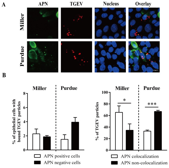
TGEV binds to APN positive/negative enterocytes. Primary enterocytes were inoculated at 4 °C with TGEV particles (m.o.i. = 10). (A) Double immunofluorescence staining of TGEV particles bound to APN positive/negative cells. Scale bar: 10 µm. (B) The percentage of cells with bound virus particles (left panel). The percentage of APN colocalized TGEV particles was counted based on five random fields (right panel). Data are expressed as the mean ± SD of the results of three separate experiments. Statistically significant differences are indicated as *p < 0.05 and ***p < 0.001.

