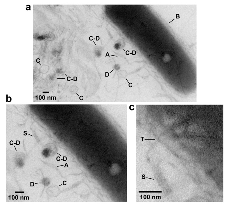
Figure 3.
EM-TS of a phage G plaque. The procedure of reference 28 was used to embed, thin section and perform electron microcopy of a plaque of phage G. (a) A field at relatively low magnification, (b) a region within the same field at higher magnification, (c) a region of the field of (b) at higher magnification and with increase in contrast. A, agarose fiber; C, capsid; D, condensed DNA; S, tail sheath; T, tail tube; B, bacterial cell.

