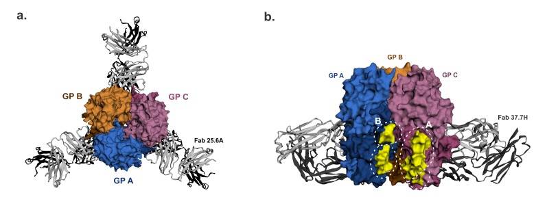
Figure 3.
GPC-B† quaternary structure epitope. The 25.6A and 37.7H monoclonal antibodies (mAbs) were raised to the LASV GPC-B conformational epitope. (a). (Top view) The LASV GP trimer is bound by three 25.6A Fabs, with each Fab binding two GP monomers near the trimer’s base. (b). (Front view rotated 60°) The GP trimer bound near its base by three 37.7H Fabs (two Fab in view) in a similar manner as above. Amino acid residues in the 37.7H epitope sites A and B highlighted in yellow to show the antibody footprint. GP monomers are shown as surface representations. LASV GP-mAb bound structures were retrieved from the Protein Database using PDB IDs 6P95 and 5VK2 for 25.6A mAb-bound GP and 37.7H mAb-bound GP respectively, image generated in EzMol 2.1 online program [60] and labelled manually.

