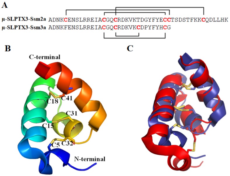
Figure 3.
(A) The sequences of µ-SLPTX3-Ssm2a and µ-SLPTX3-Ssm3a. (B) The structure of µ-SLPTX3-Ssm2a (PDB ID: 2MUN [66]). The cysteine pairs forming disulfide bonds are labeled and shown in stick representation. (C) Overlay of µ-SLPTX3-Ssm2a (dark blue) and Ta1a (red, PDB ID: 2KSL [66]). The disulfide bonds are shown in stick representation.

