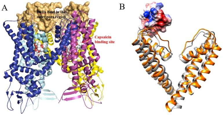
Figure 11.
The structure of polymodal transient receptor potential vanilloid 1 (TRPV1). (A) The crystal structure of TRPV1 tetramer binding with the spider peptide toxin DkTx. The cartoon is colored by TRPV1 subunits. The molecular surface in orange represents DkTx, which is bound to the outer pore region. (B) A docking model of TRPV1 monomer (orange ribbon) bound with centipede toxin RhTx (surface) by Yang et al. [49]. TRPV1 in the closed state (grey ribbon) was overlaid to the model.

