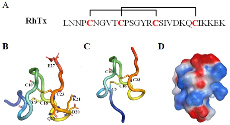
Figure 12.
(A) The sequence of RhTx. The cysteine pairs forming disulfide bonds are connected with square brackets. (B) and (C) Two representative conformations of RhTx (PDB ID: 2MVA). The key residues are labeled and shown in stick representation. (D) The electrostatic surface of RhTx. The positive and negative electrostatic potential was shown in blue and red, respectively.

