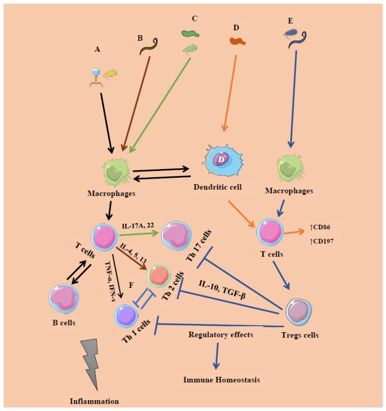
Figure 3.
The known microbe–immune interactions in inflammation and homeostasis. A. Certain members of bacteria (intracellular, systemic commensals, Proteobacteria, and opportunist pathogens) and viruses induce inflammatory responses (IR 1) by promoting T cells and their subsets T helper 1 cells (Th 1) releasing pro-inflammatory cytokines such as interferon-gamma (IFN-γ), tumor necrosis factor-alpha (TNF-α), etc., initiated by microbe-associated molecular patterns (Black arrow). B–C. Segmented filamentous bacteria (extracellular), member of Fungi, and helminths (GATA 3) induce inflammatory responses initiated at the mucosal sites, which promote the expansion of T cells expressing Th 17 and Th 2 cells releasing Interleukins (IL) 17A, IL-22, and IFN-γ, respectively (Brown and green). D. Certain members of Archaea (Methanomassiliicoccus luminyensis, Methanosphaera stadtmanae and M. smithii) promote surface markers CD86, CD197 expressed on T cells releasing Monocyte-derived dendritic cells (MODC), type 1 IFN (Orange). All the factors involving A–D may lead to tissue damage and ultimately inflammation. E–F. Certain members of Clostridia, Bacteroides fragilis, archaea, and helminths induce regulatory responses by promoting Foxp3-expressing T regulatory (Tregs) cells, limiting the activation of Th1, Th2, and Th17 cells (Blue). This regulation and tolerance promote homeostasis.

