Figure 1.
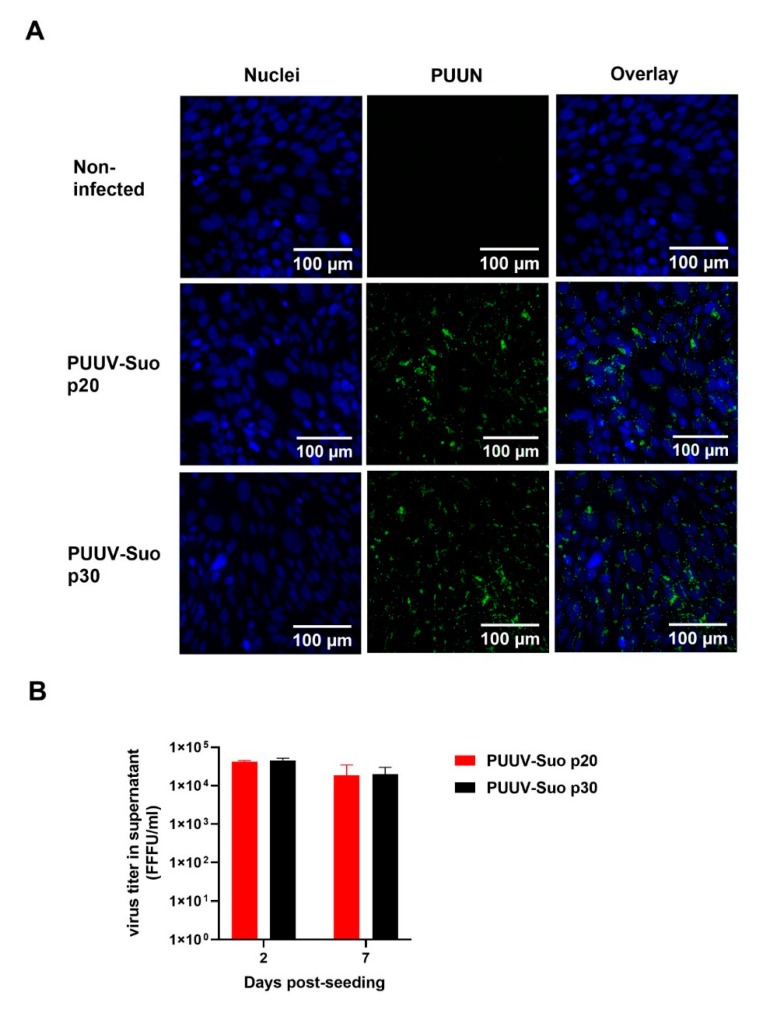
Successful isolation of PUUV-Suo in a bank vole cell line. (A) Non-infected or PUUV-Suo infected bank vole kidney epithelial cells (Mygla.REC.B) were stained for nuclei (DNA) using Hoechst 33420 (blue) and for PUUV nucleocapsid protein (PUUN) using PUUN-specific rabbit polyclonal antibody followed by AlexaFluor488-conjugated secondary antibody (green) followed by confocal microscopy analysis. PUUV-Suo infected cells at cell passages 20 or 30 post exposure to wild PUUV containing lung homogenates are shown. Images are obtained from cells grown for 2 days post-seeding. (B) Virus titers were measured in supernatants of PUUV-Suo infected Mygla.REC.B cells (passages 20 and 30) at 2 and 7 days post-seeding. Bars represent mean +/− standard deviation (n = 2).

