Figure 2.
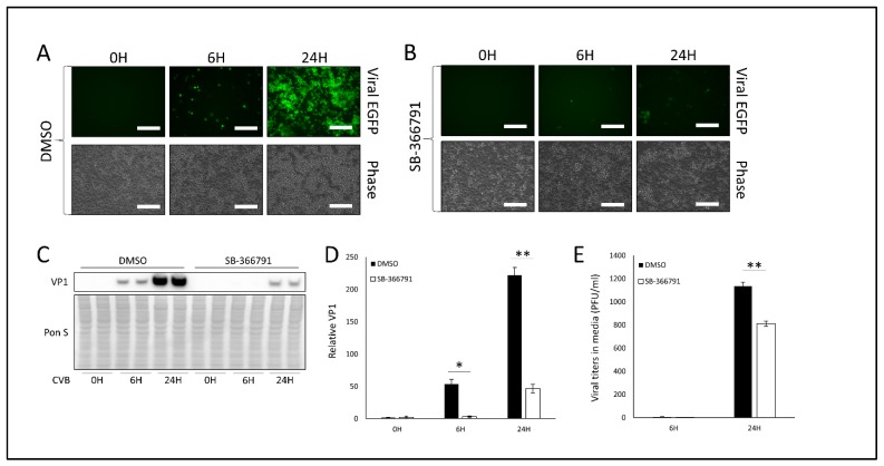
Treatment with TRPV1 inhibitor SB-366791 attenuates CVB infection- HeLa cells were treated with 10 µM of TRPV1 inhibitor SB-366791 or equivalent volume DMSO for 24 h and subsequently infected with EGFP-CVB at MOI 0.1 for 24 h. Fluorescence microscopy compares viral EGFP expression between cells treated with (A) DMSO or (B) SB-366791. Phase contrast images are shown below respective fluorescence image fields. Scale bars represent 150 µm. (C) Western blot on cell lysates from A and B detecting VP1 viral capsid protein. Ponceau S is shown below. (D) Densitometric quantification of western blot in C. (** p < 0.01; Student’s t-test, n = 3). (E) Plaque assay quantification of infectious virus in media from cells in A and B. (** p < 0.01; Student’s t-test, n = 3).

