Figure 3.
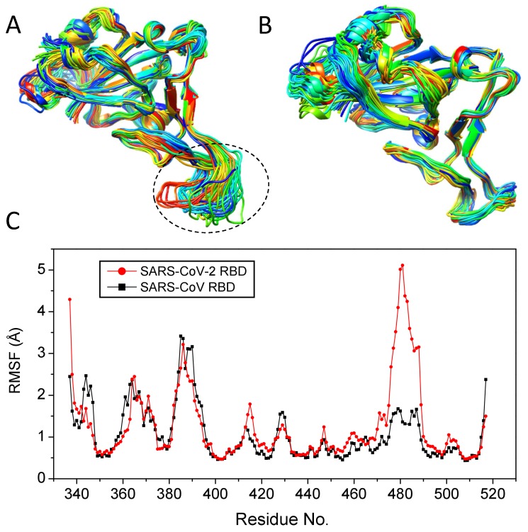
The conformational flexibility of the RBD protein. (A) The 50 representative trajectories of the SARS-CoV-2 RBD protein over a 10 ns MD simulation, where the highly flexible region (residues 470–490) is indicated by a circle. (B) The 50 representative trajectories of the SARS-CoV RBD protein over a 10 ns MD simulation. (C) The RMSFs for the RBD proteins of SARS-CoV-2 and SARS-CoV, where the residue numbering is taken from the SARS-CoV-2 RBD and the data of SARS-CoV are then aligned to those of SARS-CoV-2 according to the sequence alignment.

