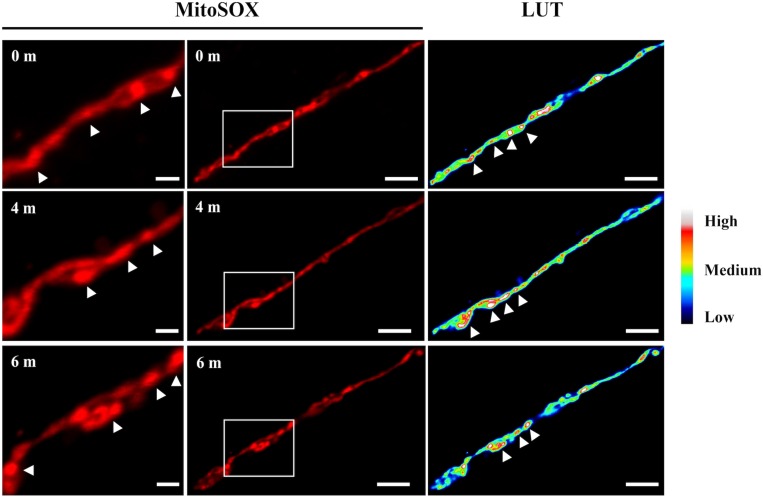
FIGURE 11.
Time course observation of ΔdnmA mitochondria stained with MitoSOX. Mycelia from strain TVG1 (ΔdnmA) grown for 14 h in supplemented glucose minimal medium was stained with MitoSOX, as indicated in section “Materials and Methods,” and then illuminated at 0, 4 and 6 min and observed by using confocal microscopy (center panels). Right panels show the relative quantification of the fluorescence intensity (LUT). Left panels show a magnification of squared regions in the central panels, where higher fluorescence intensity regions are indicated by white arrowheads. White bars = 5 μm.

