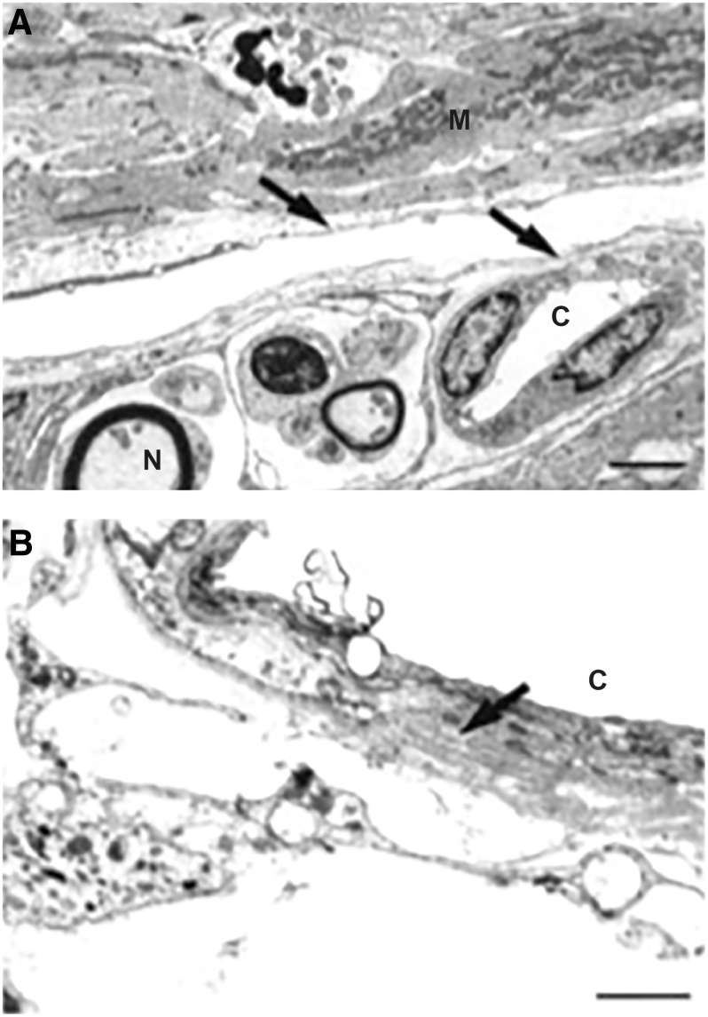
FIG. 5.
Electron micrographs of sagittal sections through the anterior longitudinal portion of the ciliary muscle of cynomolgus monkeys after 1 year of topical treatment with bimatoprost. Ciliary body remodeling after bimatoprost treatment included enlarged spaces for outflow between muscle bundles in the anterior ciliary muscle that were partially lined with endothelial-like cells (arrows in A). Capillaries within the enlarged intermuscular spaces had a thickened basement membrane (arrow in B) in contact with some endothelial-like cells. Used with permission of ARVO, from Richter et al.76; permission conveyed through Copyright Clearance Center, Inc., Scale bars: (A) 2 μm; (B) 1 μm. C, capillary; M, muscle fiber bundles; N, nerve fiber.

