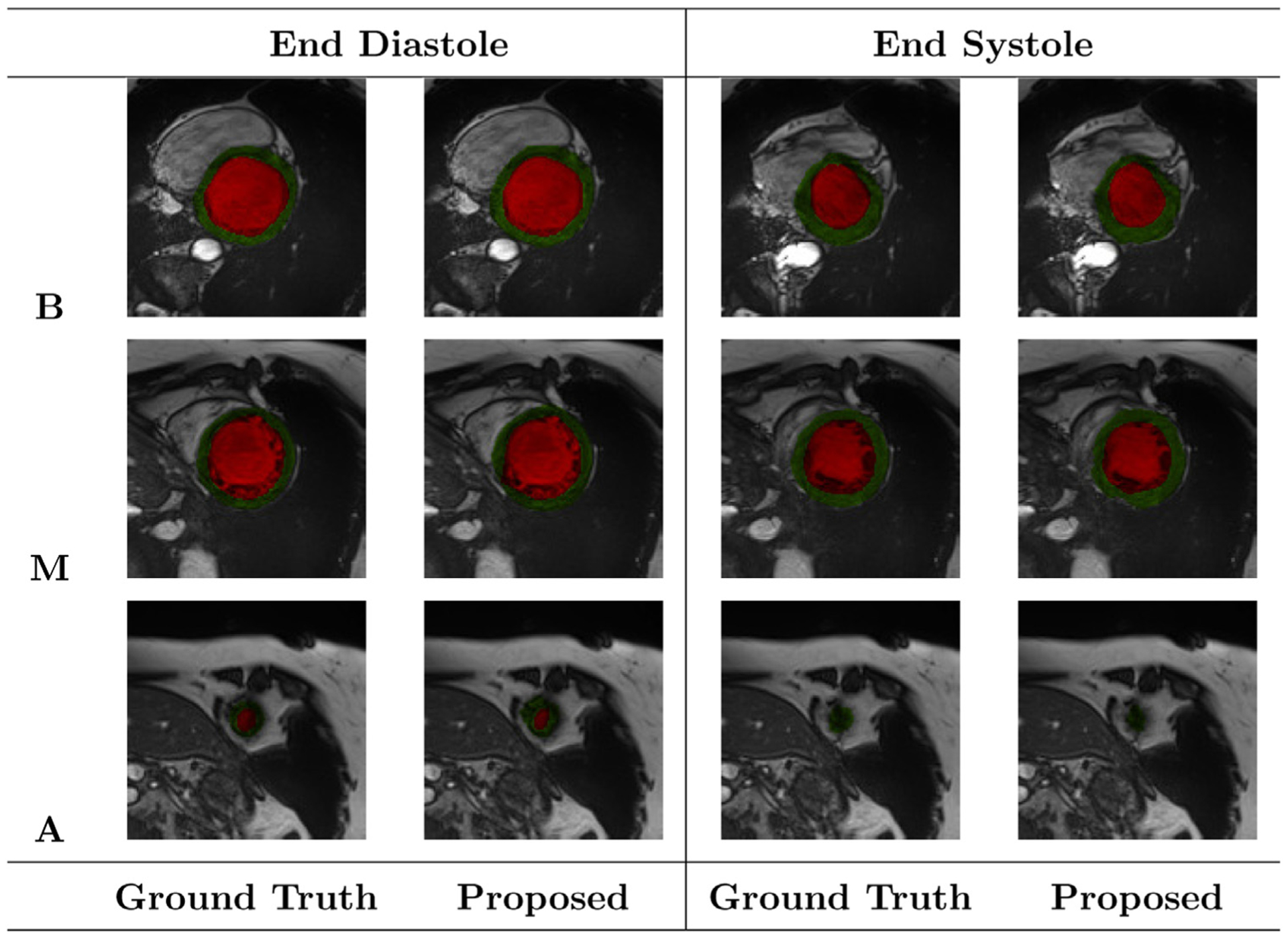
Fig. 8.

Segmentation results of the FCN2 network at ED and ES phases of one patient during a ten-fold cross-validation. Red, and green regions refer to the LV cavity, and myocardium, respectively. The letters B, M, and A refer to Basal, Mid-cavity, and Apical slices, respectively. The colors red and green indicate LV cavity and LV myocardium, respectively.
