
Figure 4.
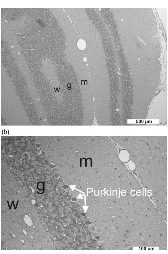
(a) The cerebellar cortex consists of three layers: the molecular layer (m), the granule cell layer (g) and the Purkinje cell layer in the middle. The white matter (w) is below the granule cell layer. Purkinje cell dendritic structures and an unmyelinated afferent fiber system, the parallel fibers, characterize the molecular layer, while the granular layer predominantly comprises small neurons supplied by myelinated afferents, the mossy fibers.55 The cranial window was implanted in the area of the cerebellar foliae V and VIa. Given the complex anatomical structure of the cerebellar foliae, the most likely explanation for the variability of the negative DC shift and the [K+]o signal during SD was that the position of the electrode tip varied with respect to the cortical layers and the white matter between different experiments. (b) Pycnotic Purkinje cells between granule cell layer and molecular layer after exposure to ET-1 at 1 µM (group 4).